


Digital Mammography Essentials
$155.00
- StudyModules
- StudyGlossary
- Post-Tests
- StudyProgress Reports
- StudyGuidelines
- Certificate of Achievement
- Targeted CE
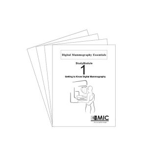
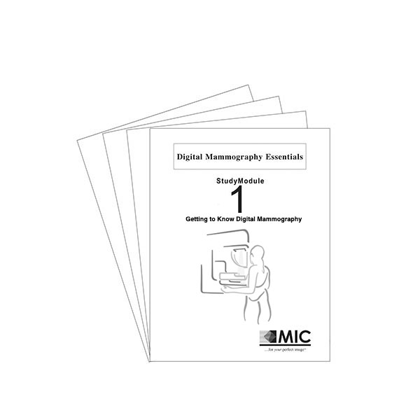
The Digital Mammography Essentials self-study course consists of 4 comprehensive StudyModules that are delivered to you all at once in a single convenient volume. Each StudyModule contains easy-to-follow text with an abundance of illustrations, clinical images and summaries, written in the language of the radiologic technologist.
- Getting to Know Digital Mammography
- Breast Cancer
- History of Mammography
- Applications of Mammography
- Mammographic Imaging Theory
- How Mammography Has Been Traditionally Performed
- Limitations of Mammography
- Sensitivity
- Specificity
- Sensitivity and Specificity Together
- Other Performance Limitations of Mammography
- Introduction of Digital Mammography
- Digital Mammography Defined
- How Digital Mammography Works: an Overview
- Benefits of Digital Information
- Early Challenges for Digital Mammography
- Technical Challenges
- Regulatory Challenges
- Market Acceptance
- Comparing Digital Mammograms to Film Mammograms
- Current State of Digital Mammography
- Performance of Digital Mammography
- Understanding a Digital Signal
- Hardware and System Operation
- Overview of a Digital Mammography System
- System Components
- Acquisition Console
- Digital-to-Analog Converter
- Introduction to the Gantry and its Functional Sections
- X-ray Generation Section of the Gantry
- High Voltage Generator
- Mammography X-ray Tube
- Beam Filtration
- Collimation
- Breast Positioning Section of the Gantry
- Compression Hardware
- Image Detection Section of the Gantry
- Anti-scatter Grid: Linear Grids, HTC Grids
- Digital Detectors: Digital Detectors vs. Screen-film, Indirect Conversion Digital, Direct Conversion Digital, Common Types of Digital Detectors
- Detector Readout Electronics
- Analog-to-Digital Converter
- Acquisition Console Image Processing
- Picture Archiving and Communication System
- Softcopy Review Workstation
- Hardcopy Device
- Image Formation and Quality
- Performing the Digital Mammography Examination
- Positioning the Breast
- Understanding Detector Field-of-View in Digital Mammography
- Exposure Techniques
- Performing the Acquisition
- Formation of a Digital Mammography Image
- How Image Pixel Values are Determined
- Displaying Digital Mammography Images
- Acquired Image vs Displayed Image
- Reviewing the Image
- Digital Mammography Image Processing: Image Processing in the Background, Interactive Image Processing
- Performing the Study: Summary
- Image Quality in Digital Mammography
- Factors That Affect Image Quality: Contrast, Resolution, Noise
- Methods Used to Characterize System Quality: Modulation Transfer Function, Detective Quantum Efficiency
- Quality Control: Detector Quality Control, AEC/Dose Performance Quality Control, Softcopy Display Quality Control, Laser Printer Quality Control
- Advanced Topics in Digital Mammography
- Computer Aided Detection
- The CAD Report
- How CAD Works
- CAD Today and into the Future
- Contrast Enhanced Mammography
- Principles of Contrast Agents
- Administration of the Contrast Agent
- Functional Imaging
- Imaging Methods
- Tomosynthesis
- Tomosynthesis Reconstruction
- Benefits and Expectations of Tomosynthesis
- Tomosynthesis in Clinical Practice
- Other “3-D” Digital Mammography Technologies
- Dedicated Breast CT Scanner
- Stereo Mammography
- Fusion Imaging
- Fusion with Ultrasound
- Fusion with Nuclear Imaging
- Fusion with Other Modalities
- Telemammography
- Breast Cancer Risk Assessment
Also included with the program is a handy StudyGlossary. The StudyGlossary contains indexed references to selected key terms in the program which include definitions as well as where to look in the StudyModules to find more information about each entry.
After you complete each StudyModule, there is a multiple choice Post-Test. Simply fill in the Post-Test Answer Sheet and return it to us using one of the following options:
- Use the postage-paid return envelope
- Fax your Answer Sheet for faster feedback
- Email it to CE@micinfo.com for the quickest results
Of course, all program information and Post-Test results are treated with the utmost confidentiality.
Each StudyModule has been separately accredited so you earn Category A credits as you pass each Post-Test, up to 13.5 with the Digital Mammography Essentials!

After we grade each Answer Sheet, we send you a StudyProgress Report that includes your Post-Test score and the official notification of the Category A CE credits earned for that Post-Test.
As you complete each Post-Test, you also receive a set of StudyGuidelines which presents detailed explanations of each and every Post-Test question! StudyGuidelines offer additional information not found in the StudyModules and provide you with guidance back to the specific section that you need to study further.
An Answer Key is provided once you achieve a score of at least 75%. If you score below 75%, an additional Answer Sheet is provided, and you are offered another chance at the Post-Test after you study the material further. StudyProgress Reports and StudyGuidelines are sent to you via first class mail, normally within two business days after we receive your Post-Test answers.

You are awarded the final Certificate of Achievement after you earn the Continuing Education credits for all the StudyModules.
Targeted CE per ARRT’s Discipline, Category, and Subcategory classification for enrollments starting after September 19, 2023:
[Note: Discipline-specific Targeted CE credits may be less than the total Category A credits approved for this course.]
StudyModule 1 [3.25 credits]:
- Mammography: 2.50
Patient Care: 0.25
Patient Interactions and Management: 0.25
Image Production: 2.00
Image Acquisition and Quality Assurance: 2.00
Procedures: 0.25
Mammographic Positioning, Special Needs, and Imaging Procedures: 0.25
StudyModule 2 [3.25 credits]:
- Mammography: 3.25
Image Production: 3.25
Image Acquisition and Quality Assurance: 3.25
StudyModule 3 [3.5 credits]:
- Mammography: 3.50
Image Production: 3.25
Image Acquisition and Quality Assurance: 3.25
Procedures: 0.25
Mammographic Positioning, Special Needs, and Imaging Procedures: 0.25
StudyModule 4 [3.5 credits]:
- Mammography: 2.00
Image Production: 1.75
Image Acquisition and Quality Assurance: 1.75
Procedures: 0.25
Mammographic Positioning, Special Needs, and Imaging Procedures: 0.25