


CT CrossTrainer
$199.00
- StudyModules
- StudyGlossary
- Post-Tests
- StudyProgress Reports
- StudyGuidelines
- Certificate of Achievement
- Targeted CE
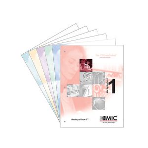
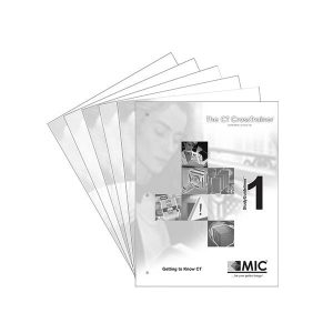
The CT CrossTrainer self-study course consists of 6 comprehensive StudyModules
that are delivered to you all at once in a handy reference binder.
Each StudyModule contains easy-to-follow text with an abundance of
illustrations, clinical images and summaries, written in the language of the radiologic technologist.
- Getting to Know CT
- History of Radiology Leading to CT
- X-ray Started it All
- Tomography was Introduced
- Computers Made it Possible
- CT Scanner Designs
- Benefits of CT
- Getting the Inside Story
- Visually Differentiating Tissues
- Digital Images
- Short Acquisition Time
- Overview of CT
- Review of X-ray Production
- Attenuation of X-rays
- Detectors Measure the X-rays
- Image Reconstruction
- Image Display and Archiving
- CT Hardware & System Operation
- The Flow of Data Through a CT System
- The Operator’s Console
- The Host Computer
- The Scan Controller
- The Digital-to-Analog Convertor
- Gantry and Table Controllers
- The High Voltage Generator
- The CT X-ray Tube
- Collimators
- Detectors
- The Amplifier and the ADC
- The Array Processor
- PACS
- Forming CT Images
- Collecting Attenuation Information
- Different Scanning Methods
- Localizer Scan
- Conventional Scan
- Helical Scan
- CT Image Reconstruction
- Back Projection
- Cone Beam Reconstruction
- Convolution
- Iterative Reconstruction
- Digital Cross-sectional Images
- The Gray Scale
- Post-Processing Techniques
- Retrospective Reconstruction
- Multiplanar Reconstruction
- 3D Surface Rendering
- CT Image Appearance
- Contrast
- Resolution
- Noise
- Dose
- mAs
- kVp
- Slice Thickness
- Table Increment
- Pitch
- Reconstruction Interval
- Field-of-View
- Matrix
- Reconstruction Filter
- Beam Hardening Artifact
- Partial Volume Artifact
- Motion Artifact
- Metal Artifact
- Windowing the CT Image
- Applying a Display Filter
- Magnifying the CT Image
- CT Safety
- Radiation Concerns
- Measuring the Harmful Effects
- Effective Dose
- Minimizing the Patient Dose
- Occupational Exposure
- Contrast Agent Concerns
- What is Contrast Media?
- Intravenous Contrast Agents
- Oral Contrast Agents
- Intrathecal Contrast Agents
- Emergency Procedures
- Code Procedures
- Vital Signs
- Special Pediatric Care
- Sedation
- Minimizing Dose
- Contrast Administration
- The CT Exam
- The Patient Experience
- Preparation
- The CT Exam
- After the CT Exam
- Typical CT Exams
- Head, Neck, Spine, Joints, Chest, Abdomen, Pelvis
- CT Angiography
- CT Perfusion
- CT Colonography
- PET/CT

Also included with the program is a pocket StudyGlossary.
The StudyGlossary contains indexed references to all key terms
in the program which include definitions as well as where to look
in the StudyModules to find more information about each entry.

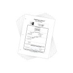
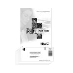
After you complete each StudyModule, there is a multiple choice Post-Test. Simply fill in the Post-Test Answer Sheet and return it to us using one of the following options:
- Use the postage-paid return envelope
- Fax your Answer Sheet for faster feedback
- Email it to CE@micinfo.com for the quickest results
Of course, all program information and Post-Test results are treated with the utmost confidentiality.
Each StudyModule has been separately accredited so you earn Category A credits as you pass each Post-Test, up to 20.75 with the CT CrossTrainer!

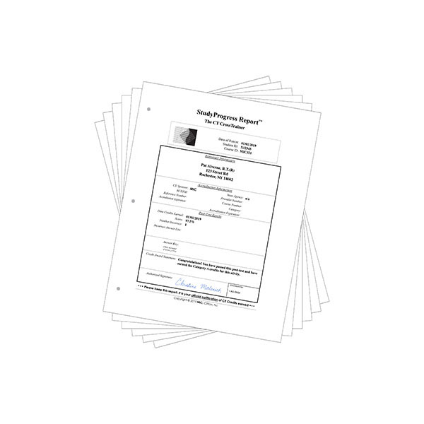
After we grade each Answer Sheet, we send you a StudyProgress Report that includes your Post-Test score and the official notification of the Category A CE credits earned for that Post-Test.
As you complete each Post-Test, you also receive a set of StudyGuidelines which presents detailed explanations of each and every Post-Test question! StudyGuidelines offer additional information not found in the StudyModules and provide you with guidance back to the specific section that you need to study further.
An Answer Key is provided once you achieve a score of at least 75%. If you score below 75%, an additional Answer Sheet is provided, and you are offered another chance at the Post-Test after you study the material further. StudyProgress Reports and StudyGuidelines are sent to you via first class mail, normally within two business days after we receive your Post-Test answers.

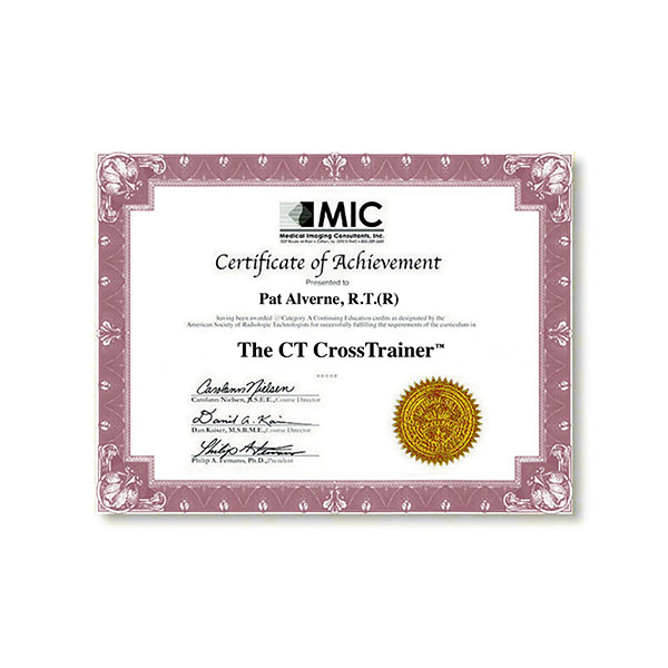
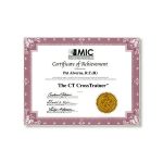
You are awarded the final Certificate of Achievement
after you earn the Continuing Education credits for
all the StudyModules.
The CT CrossTrainer fulfills ARRT’s entire 16 credit Structured Education Requirement for the Discipline of Computed Tomography.
Targeted CE per ARRT’s Discipline, Category, and Subcategory classification for enrollments starting after March 9, 2023:
[Note: Discipline-specific Targeted CE credits may be less than the total Category A credits approved for this course.]
StudyModule 1 [3.25 credits]:
- Computed Tomography: 2.50
Image Production: 2.50
Image Formation: 1.25
Image Evaluation and Archiving: 1.25
StudyModule 2 [2.75 credits]:
- Computed Tomography: 2.75
Safety: 0.50
Radiation Safety and Dose: 0.50
Image Production: 2.25
Image Formation: 2.00
Image Evaluation and Archiving: 0.25 - Nuclear Medicine Technology: 1.00
Image Production: 1.00
Instrumentation: 1.00 - Radiation Therapy: 1.00
Procedures: 1.00
Treatment Volume Localization: 1.00
StudyModule 3 [3 credits]:
- Computed Tomography: 3.00
Image Production: 3.00
Image Formation: 2.75
Image Evaluation and Archiving: 0.25 - Nuclear Medicine Technology: 1.00
Image Production: 1.00
Instrumentation: 1.00 - Radiation Therapy: 1.00
Procedures: 1.00
Treatment Volume Localization: 1.00
StudyModule 4 [3.5 credits]:
- Computed Tomography: 3.50
Safety: 0.25
Radiation Safety and Dose: 0.25
Image Production: 3.25
Image Formation: 1.50
Image Evaluation and Archiving: 1.75 - Nuclear Medicine Technology: 1.00
Image Production: 1.00
Instrumentation: 1.00 - Radiation Therapy: 1.00
Procedures: 1.00
Treatment Volume Localization: 1.00
StudyModule 5 [3.75 credits]:
- Computed Tomography: 3.75
Patient Care: 2.25
Patient Interactions and Management: 2.25
Safety: 1.50
Radiation Safety and Dose: 1.50 - Registered Radiologist Assistant: 3.50
Patient Care: 2.25
Patient Management: 0.25
Pharmacology: 2.00
Safety: 1.25
Patient Safety, Radiation Protection, and Equipment Operation: 1.25 - Radiation Therapy: 0.75
Patient Care: 0.75
Patient Interactions and Management: 0.75
StudyModule 6 [4.5 credits]:
- Computed Tomography: 4.50
Patient Care: 0.50
Patient Interactions and Management: 0.50
Procedures: 4.00
Head, Spine, and Musculoskeletal: 2.00
Neck and Chest: 1.00
Abdomen and Pelvis: 1.00 - Nuclear Medicine Technology: 0.25
Procedures: 0.25
Other Imaging Procedures: 0.25 - Registered Radiologist Assistant: 0.50
Patient Care: 0.50
Pharmacology: 0.50 - Radiation Therapy: 0.50
Patient Care: 0.50
Patient Interactions and Management: 0.50